Mohs Surgery
Mohs micrographic surgery — the highest cure rate for many skin cancers with maximal healthy-tissue preservation — performed by Dr. Blair Harris in Leland, NC.
About Mohs Surgery
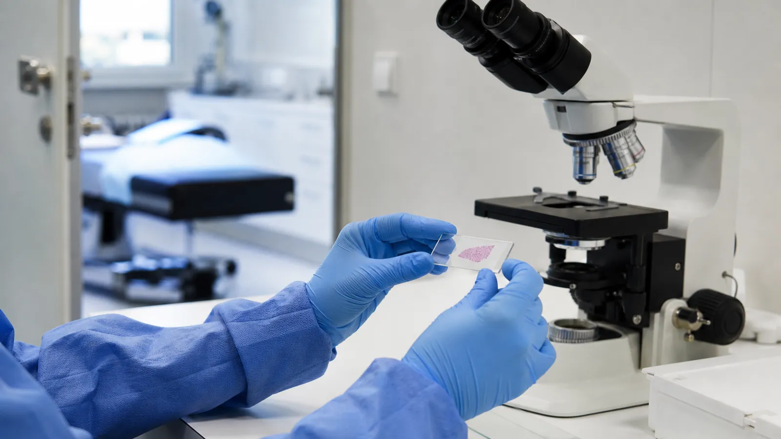
Mohs micrographic surgery removes skin cancer in thin layers, with each layer examined under the microscope while you wait — so surgery ends the moment the margins are clear, and not a millimeter more healthy tissue is taken than necessary. That precision gives Mohs the highest cure rates for basal cell and squamous cell carcinoma and makes it the treatment of choice for cancers on the face, ears, nose, lips, and hands, where tissue preservation matters most. Dr. Blair Harris, a double board-certified dermatologist and Mohs surgeon, performs the entire procedure — surgery, microscopic examination, and reconstruction — in our Leland office.
Treatment Options
Mohs surgery offers several advantages, including a high cure rate, maximal preservation of healthy tissue, and the immediate examination of removed tissue during the procedure. It is particularly beneficial for treating skin cancers on sensitive areas such as the face, where tissue preservation is crucial for both functional and cosmetic reasons. However, Mohs surgery may not be suitable for all types of skin cancer or all patients, and the decision to use this technique depends on various factors, including the type and location of the cancer.
Mohs Procedure Steps
The systematic process of Mohs micrographic surgery performed by Dr. Harris.
Types of Mohs Procedure Steps
Local Anesthesia
The patient is administered local anesthesia to numb the area where the surgery will take place. This ensures that the patient remains comfortable throughout the procedure.
Tissue Removal
Dr. Harris starts by removing a thin layer of the cancerous tissue. This layer is then divided into sections and labeled for precise mapping.
Tissue Examination
The removed tissue sections are immediately frozen, prepared, and examined under a microscope by Dr. Harris. This allows for the immediate detection of cancerous cells.
Mapping and Additional Layers
If cancer cells are still present in the examined tissue, Dr. Harris uses the mapped sections to precisely locate the affected areas. Additional layers of tissue are then removed only from those specific areas, sparing as much healthy tissue as possible.
Complete Removal
The process is repeated until no cancer cells are detected in the examined tissue, indicating that the entire tumor has been removed. This ensures a high cure rate and minimizes the removal of healthy tissue.
Closure
Once the cancerous tissue has been completely excised, Dr. Harris discusses the options for closing the wound. Depending on the size and location of the wound, various closure techniques such as secondary intention, sutures, skin grafts, or flaps may be employed.
Ready to get started?
Schedule a consultation with our board-certified dermatologist.
Treatment Results
Medical Graphic Content Warning
The following photos depict surgical incisions, stitches, and post-operative wounds. Each image is blurred by default. Click “Show image” on any photo to reveal it, or “Show all” above. Once revealed, click the photo or “Enlarge” to view it full size.

Graphic medical content — surgical incisions and stitches
Case 1 — Pre-operative markings, post-operative closure, and healed result

Graphic medical content — surgical incisions and stitches
Case 2 — Pre-operative markings, post-operative closure, and healed result

Graphic medical content — surgical incisions and stitches
Case 3 — Pre-operative markings, post-operative closure, and healed result

Graphic medical content — surgical incisions and stitches
Case 4 — Pre-operative markings, post-operative closure, and healed result

Graphic medical content — surgical incisions and stitches
Case 5 — Pre-operative markings, post-operative closure, and healed result

Graphic medical content — surgical incisions and stitches
Case 6 — Pre-operative markings, post-operative closure, and healed result

Graphic medical content — surgical incisions and stitches
Case 7 — Pre-operative markings, post-operative closure, and healed result

Graphic medical content — surgical incisions and stitches
Case 8 — Pre-operative markings, post-operative closure, and healed result

Graphic medical content — surgical incisions and stitches
Case 9 — Pre-operative markings, post-operative closure, and healed result

Graphic medical content — surgical incisions and stitches
Case 10 — Pre-operative markings, post-operative closure, and healed result
Individual results may vary. Photos are for informational purposes only and may not reflect typical results.
Pre & Post Care
Pre-Treatment
- Discuss medications that may affect bleeding
- Avoid aspirin and blood thinners as directed
- Plan for the length of the procedure (several hours)
- Arrange transportation as you may need to return for multiple stages
- Follow preoperative fasting instructions if sedation is used
Post-Treatment
- Keep the surgical site clean and protected
- Follow wound care instructions carefully
- Attend all follow-up appointments
- Avoid strenuous activities that could disrupt healing
- Use sun protection on the treated area
- Monitor for signs of infection or complications
Expected Results
Onset
Complete cancer removal during the procedure with immediate microscopic verification
Duration
Wound healing takes 2-6 weeks depending on closure method and location
Maintenance
Regular skin cancer surveillance and follow-up care, typically every 6-12 months
Ongoing Care
Plan to spend a good part of the day with us: most of the visit is waiting while each tissue layer is processed and examined, and most cancers clear within one to three stages. Once margins are clear, Dr. Harris repairs the site the same day — our before-and-after gallery shows how well these repairs can heal. Follow-up skin exams afterward are important, since having one skin cancer increases the likelihood of developing others.
Questions about Mohs Surgery?
We're happy to help you decide the best next step.
Related Services
Skin Biopsy
Learn more about this service
Learn moreSkin Cancer Removal
Learn more about this service
Learn moreScar Revision
Learn more about this service
Learn moreMole Removal
Learn more about this service
Learn morePsoriasis
Learn more about this service
Learn moreAtopic Dermatitis (Eczema)
Learn more about this service
Learn moreYour Physician
About Blair Harris, DO, FAAD
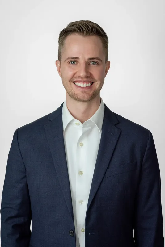
Blair Harris, DO, FAAD
Double Board-Certified Dermatologist and Mohs Surgeon
Dr. Harris brings extensive expertise in both general and surgical dermatology, providing comprehensive skin care for patients in the Leland area. With double board certification and specialized training in Mohs surgery, he delivers the highest standard of care for skin cancer treatment and complex dermatological conditions.